
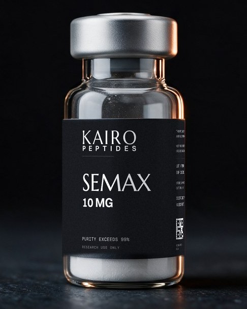
Semax
10MGStudied for memory and learning, neuroprotection, recovery after a loss of blood flow in the brain, and stress resilience. Every paper behind those is linked below.
$45.00
- PURITY EXCEEDS 99%
- COA WITH EVERY BATCH
- 3RD-PARTY VERIFIED
| Title | Range | Discount |
|---|---|---|
| Pack tier: Semax (KAIRO-SEMAX-10MG) | 1 - 2 | $45.00 |
| Pack of 3+: 10% off | 3 - 4 | $40.50 |
| Pack of 5+: 15% off | 5 - 9 | $38.25 |
| Pack of 10+: 20% off | 10 + | $36.00 |
For laboratory research use only. Not for human or veterinary use, not for diagnostic or therapeutic use, and not for food or drug manufacture. No preparation or usage guidance is provided anywhere on this page.
Memory and learning. Nerve cells under stress.
That is what almost all of this research measures, nearly always in rats, and it is why Semax keeps turning up in learning research and brain injury research at the same time.
-
Memory and learning
Preclinical preliminary A single study, an early mechanistic observation, or a result whose interpretation is the authors' own inference.Semax is studied for learned behavior in animals, reported to increase a learned avoidance response in rats and to improve learning measures in rat pups. Nothing on this page measured memory or learning in a person.
BDNF and TrkB neurotrophin signaling in hippocampus and basal forebrain Early-life drug exposure model
-
Neuroprotection
Preclinical moderate A well-described result with several independent outcome measures, but from a single laboratory or a single study design.Semax is studied for how brain tissue holds up after the blood supply to it is blocked. In rats it was reported to lower damage and inflammation proteins next to the injury, to leave more nerve cells alive in the tissue around it, and to reduce the visible signs of nerve tissue damage.
Cerebral ischemia models: specific proteins and tissue appearance Cerebral ischemia models: blood vessels and cell multiplication around the injury
-
Nerve growth and connections
Preclinical moderate A well-described result with several independent outcome measures, but from a single laboratory or a single study design.Semax is studied for nerve cell growth and new connections through its effect on BDNF, a protein that helps nerve cells survive and rewire. It was reported to raise BDNF in the rat hippocampus after a single nose-drop application, by 1.4-fold at most, far below the numbers this market repeats.
BDNF and TrkB neurotrophin signaling in hippocampus and basal forebrain Neurotrophin gene switching in cultured support cells
-
Recovery after a loss of blood flow in the brain
Preclinical moderate A well-described result with several independent outcome measures, but from a single laboratory or a single study design.Semax is studied in rats whose blood supply to the brain was blocked, and in 110 people in rehabilitation after the same kind of event. In those 110 it tracked with higher BDNF in the blood and better recovery scores, in a study that describes no random assignment, so what it shows is an association and not a cause.
Cerebral ischemia models: which genes get switched on and off Human study: recovery after a loss of blood flow in the brain, and a blood growth-factor marker
-
Serotonin and dopamine chemistry
Preclinical moderate A well-described result with several independent outcome measures, but from a single laboratory or a single study design.Semax is studied for serotonin and dopamine chemistry in the striatum, a deep brain region. It was reported to raise a serotonin breakdown product in rodents, while dopamine itself did not move at all in that same study, which is worth knowing because much of this market says it does.
-
Stress resilience
Preclinical moderate A well-described result with several independent outcome measures, but from a single laboratory or a single study design.Semax is studied under stress in rats, reported to blunt the behavior changes an acute stressor caused. Given across weeks of unpredictable stress it was reported to restore a normal preference for sugar water and normal BDNF in the hippocampus, while a forced swim measure in the same study did not move.
Every line above describes what was measured in a laboratory. None of it establishes what the compound would do in a person.
Frequently paired with
What shoppers usually add alongside this one
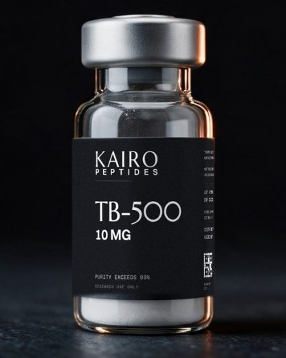
A versatile peptide, studied in research on cell movement and tissue repair.
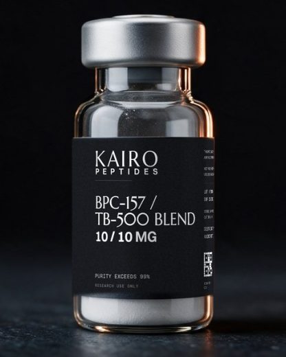
A recovery-focused blend, studied in research on tissue repair.
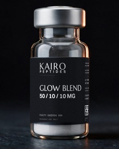
A multi-peptide skin blend, studied in research on skin and tissue.
What the research says about pairing these
Pro-Gly-Pro (PGP), this compound's own tail fragment
The best-sourced pairing in this literature, and the only one that is genuinely a research relationship rather than a retail one. Pro-Gly-Pro is a documented breakdown product of the parent peptide, and several rat ischemia studies deliberately run the two as parallel arms in order to separate the parent peptide's effects from its fragment's. It is a mechanistic dissection, not a stack, and the two are not given together in any of it.
The parallel-arm studies report divergence as often as agreement, which is the scientifically interesting part. PMID 19633950 reported that Semax acted selectively in ischemic cortex while the fragment's influence was mainly non-specific. PMID 20617398 reported that both increased the multiplication of support cells, vessel lining and progenitor cells, but that only Semax reduced visible ischemic damage. PMID 22295573 reported opposite directions of receptor gene-message change in frontal cortex for the two. PMID 18239779 tested both against glutamate toxicity in a dish. PMID 36553646 reported that Pro-Gly-Pro and Pro-Gly-Pro-Leu showed largely Semax-unlike effects on inflammatory and nerve-signaling genes after a temporary artery blockage. No study gives the two together.
ACTH(6-9)PGP and related melanocortin analogs
Comparative preclinical work runs Semax alongside other ACTH-derived peptides in the same rat models, to map the shared mechanism and compare structures. These are separate arms in comparative studies, not combinations, and none of these analogs is sold here.
PMID 39767736 reported that both peptides significantly reduced the ischemia-caused distortion in expression for 1171 genes at 24 hours after a temporary artery blockage. PMID 40650034 compared their effects across brain regions with different degrees of ischemic damage and reported that the two regions responded differently. PMID 37510287 compared them in the early window after the blockage. PMID 38534749 examined both for blood-vessel and support-cell outcomes. PMID 34576218 compared them under acute restraint stress.
Selank
Defensible to mention only with precise framing. The two peptides have been examined side by side inside the same published protocols, always as separate arms, and they are sold alongside each other in this market. The co-studied flag above is off deliberately: appearing in the same paper is not the same thing as being given together, and a two-word badge cannot carry that distinction, so it is stated here instead.
PMID 11443939 tested both against enkephalin-degrading enzymes in human serum and reported Semax the more potent. PMID 32342318 scanned 52 healthy participants receiving Semax, Selank, or placebo in one MRI study and reported both shared and compound-specific connectivity effects. PMID 28702721 tested both in rats with a chemically induced injury to the brain's dopamine system, where neither changed motor activity and only Selank reduced anxiety measures. In every one of those cases the two were separate arms and were never given together. No published study located in the verification pass has evaluated the two in combination, so any complementary-mechanism story is a hypothesis assembled from two separate literatures rather than a demonstrated result.
BPC-157 and TB-500
Frequently co-marketed in retail peptide bundles as a recovery-plus-cognition pairing. That is an observed market pattern and nothing more.
No published study located in the verification pass gives Semax with either compound, and no shared mechanism has been demonstrated. This must never be presented as a research-supported combination.
Noopept, Cerebrolysin, racetams and other nootropics
Co-marketed and discussed in community forums as cognition stacks. Not research-supported.
No controlled study of any of these in combination with Semax was located in the verification pass.
-
Studied for
BDNF and TrkB neurotrophin signaling in hippocampus and basal forebrain
In rats, a single nose-drop application of Semax was reported to raise the level of BDNF, a protein that supports the survival and plasticity of nerve cells, in the hippocampus, along with a rise in the activation of TrkB, the receptor BDNF acts through. The reported changes were modest: BDNF protein rose at most 1.4-fold and TrkB activation 1.6-fold, with the corresponding gene messages rising about 3-fold and 2-fold. The animals given the peptide also performed more of a learned avoidance response, and the authors proposed the BDNF/TrkB system as the pathway responsible. A companion paper from the same group reported that a radioactively tagged version of the peptide bound specifically, reversibly and calcium-dependently to membranes prepared from rat basal forebrain tissue, and that BDNF protein rose in the basal forebrain three hours after application but did not rise in the cerebellum. The magnitudes matter here: these are single-digit fold changes in one brain region of a rat, not the large multiples that circulate in promotional writing. in rat studies (nose-drop application to live rats, plus a binding assay on membranes isolated from rat brain tissue) Preclinical moderate A well-described result with several independent outcome measures, but from a single laboratory or a single study design. Open for the full finding and the paper it came from.In the literatureBDNF and TrkB neurotrophin signaling in hippocampus and basal forebrain
In rats, a single nose-drop application of Semax was reported to raise the level of BDNF, a protein that supports the survival and plasticity of nerve cells, in the hippocampus, along with a rise in the activation of TrkB, the receptor BDNF acts through. The reported changes were modest: BDNF protein rose at most 1.4-fold and TrkB activation 1.6-fold, with the corresponding gene messages rising about 3-fold and 2-fold. The animals given the peptide also performed more of a learned avoidance response, and the authors proposed the BDNF/TrkB system as the pathway responsible. A companion paper from the same group reported that a radioactively tagged version of the peptide bound specifically, reversibly and calcium-dependently to membranes prepared from rat basal forebrain tissue, and that BDNF protein rose in the basal forebrain three hours after application but did not rise in the cerebellum. The magnitudes matter here: these are single-digit fold changes in one brain region of a rat, not the large multiples that circulate in promotional writing.References 1, 2
Modelin rat studies (nose-drop application to live rats, plus a binding assay on membranes isolated from rat brain tissue)
-
Studied for
Cerebral ischemia models: which genes get switched on and off
Rats given an experimental blockage of a cerebral artery were used to read out, across the whole genome, which genes change and whether the peptide changes them back. The direction of the reported effect depends on which model was used, so the two datasets should not be merged. In a model where blood flow is blocked and then restored, sequencing found 394 genes reading differently at 24 hours, with inflammation genes turned down and nerve-signaling genes turned up, the mirror image of what the blockage alone did. In a model where the blockage is permanent, a whole-genome chip study reported that the peptide mostly increased immune-system gene activity, which made up more than half of all the genes it changed at 24 hours, along with changes in blood-vessel genes. A third study reported that both Semax and its own Pro-Gly-Pro fragment increased the gene messages for growth factors and their receptors in the cortex after a permanent blockage, with Semax acting selectively in the injured cortex while the fragment's influence was mostly non-specific. This is the deepest mechanistic dataset that exists on this compound, and it is entirely in rats. in rat studies (two different surgical models: temporary artery blockage with restored blood flow, and permanent artery blockage) Preclinical moderate A well-described result with several independent outcome measures, but from a single laboratory or a single study design. Open for the full finding and the paper it came from.In the literatureCerebral ischemia models: which genes get switched on and off
Rats given an experimental blockage of a cerebral artery were used to read out, across the whole genome, which genes change and whether the peptide changes them back. The direction of the reported effect depends on which model was used, so the two datasets should not be merged. In a model where blood flow is blocked and then restored, sequencing found 394 genes reading differently at 24 hours, with inflammation genes turned down and nerve-signaling genes turned up, the mirror image of what the blockage alone did. In a model where the blockage is permanent, a whole-genome chip study reported that the peptide mostly increased immune-system gene activity, which made up more than half of all the genes it changed at 24 hours, along with changes in blood-vessel genes. A third study reported that both Semax and its own Pro-Gly-Pro fragment increased the gene messages for growth factors and their receptors in the cortex after a permanent blockage, with Semax acting selectively in the injured cortex while the fragment's influence was mostly non-specific. This is the deepest mechanistic dataset that exists on this compound, and it is entirely in rats.References 3, 4, 5
Modelin rat studies (two different surgical models: temporary artery blockage with restored blood flow, and permanent artery blockage)
-
Studied for
Cerebral ischemia models: specific proteins and tissue appearance
A rat study that measured four pre-chosen proteins after a temporary artery blockage reported that, at 24 hours, an activated form of CREB (a switch associated with repair) was higher in deeper brain structures including the damaged area, while MMP-9 and c-Fos (both associated with damage and inflammation) were lower in the neighboring cortex, and activated JNK was lower in both tissues. This study looked at four proteins picked in advance, not at the proteins of the brain as a whole. Separately, a pilot study that examined brain tissue under the microscope after a different injury, a partial whole-brain loss of blood flow, reported that both Semax and its Pro-Gly-Pro fragment increased the multiplication of support cells, blood-vessel lining cells and progenitor cells, but that only Semax reduced the visible signs of damage to nerve tissue. in rat studies (temporary artery-blockage model for the protein study; a partial whole-brain loss of blood flow for the tissue study, which its own authors call a pilot) Preclinical moderate A well-described result with several independent outcome measures, but from a single laboratory or a single study design. Open for the full finding and the paper it came from.In the literatureCerebral ischemia models: specific proteins and tissue appearance
A rat study that measured four pre-chosen proteins after a temporary artery blockage reported that, at 24 hours, an activated form of CREB (a switch associated with repair) was higher in deeper brain structures including the damaged area, while MMP-9 and c-Fos (both associated with damage and inflammation) were lower in the neighboring cortex, and activated JNK was lower in both tissues. This study looked at four proteins picked in advance, not at the proteins of the brain as a whole. Separately, a pilot study that examined brain tissue under the microscope after a different injury, a partial whole-brain loss of blood flow, reported that both Semax and its Pro-Gly-Pro fragment increased the multiplication of support cells, blood-vessel lining cells and progenitor cells, but that only Semax reduced the visible signs of damage to nerve tissue.References 6, 7
Modelin rat studies (temporary artery-blockage model for the protein study; a partial whole-brain loss of blood flow for the tissue study, which its own authors call a pilot)
-
Studied for
Cerebral ischemia models: blood vessels and cell multiplication around the injury
In rats given a temporary artery blockage, two related peptides (Semax and a close relative) were reported, 24 hours later, to leave a higher density of nerve cells in the rim of tissue around the infarct than in rats given saline, to increase the number of dividing support cells both in that rim and in undamaged cortex, and, by staining for a blood-vessel marker, to increase both the density and the size of blood vessels in that rim. All three readouts come from one study at one timepoint. in rat studies (temporary artery-blockage model, tissue staining at 24 hours) Preclinical moderate A well-described result with several independent outcome measures, but from a single laboratory or a single study design. Open for the full finding and the paper it came from.In the literatureCerebral ischemia models: blood vessels and cell multiplication around the injury
In rats given a temporary artery blockage, two related peptides (Semax and a close relative) were reported, 24 hours later, to leave a higher density of nerve cells in the rim of tissue around the infarct than in rats given saline, to increase the number of dividing support cells both in that rim and in undamaged cortex, and, by staining for a blood-vessel marker, to increase both the density and the size of blood vessels in that rim. All three readouts come from one study at one timepoint.References 8
Modelin rat studies (temporary artery-blockage model, tissue staining at 24 hours)
-
Studied for
Serotonin and dopamine chemistry in the striatum
In rodents, Semax was reported to raise a breakdown product of serotonin (5-HIAA) in the striatum, a deep brain region, by about 25 percent in tissue at two hours and by up to about 180 percent in the fluid around the cells over one to four hours, which the authors read as a positive modulating effect on the striatal serotonin system. In the same study the peptide on its own did not change dopamine or its breakdown products at all; extra dopamine release and extra movement appeared only when the peptide was given before amphetamine, which makes that a boost of the stimulant's effect rather than an effect of the peptide by itself. A second study in white rats with a chemically induced dopamine-system injury reported that the peptide reduced the severity of the resulting behavioral disturbances. A third study, using a different chemical injury model of the brain's dopamine system in rats, reported a null result for Semax: neither Semax nor Selank changed motor activity in a maze or a passive-defense test, and only Selank reduced anxiety measures. RODENT in rodent studies (the serotonin and dopamine chemistry paper says only 'in rodents' and does not name the species; the two lesion-model papers are both explicitly in rats) Preclinical moderate A well-described result with several independent outcome measures, but from a single laboratory or a single study design. Open for the full finding and the paper it came from.In the literatureSerotonin and dopamine chemistry in the striatum
In rodents, Semax was reported to raise a breakdown product of serotonin (5-HIAA) in the striatum, a deep brain region, by about 25 percent in tissue at two hours and by up to about 180 percent in the fluid around the cells over one to four hours, which the authors read as a positive modulating effect on the striatal serotonin system. In the same study the peptide on its own did not change dopamine or its breakdown products at all; extra dopamine release and extra movement appeared only when the peptide was given before amphetamine, which makes that a boost of the stimulant's effect rather than an effect of the peptide by itself. A second study in white rats with a chemically induced dopamine-system injury reported that the peptide reduced the severity of the resulting behavioral disturbances. A third study, using a different chemical injury model of the brain's dopamine system in rats, reported a null result for Semax: neither Semax nor Selank changed motor activity in a maze or a passive-defense test, and only Selank reduced anxiety measures.References 9, 10, 11
Modelin rodent studies (the serotonin and dopamine chemistry paper says only 'in rodents' and does not name the species; the two lesion-model papers are both explicitly in rats)
-
Studied for
Copper binding and the chemistry of the amyloid-beta protein
Test-tube and cell work reported that Semax forms a stable complex with copper, prevents the amyloid-beta protein from forming its own complex with copper, and shows anti-clumping and protective properties particularly when copper is present, with the authors proposing that it blocks fiber formation by interfering with the copper-bound form of the protein. A follow-up reported that the peptide can strip copper away from copper-loaded amyloid-beta, change how that complex cycles electrons, lower the reactive oxygen species it generates, and protect a human nerve-like cell line from the resulting oxidative stress. This is chemistry and cell-dish work. It is not evidence about any condition in a person. in test tubes and cultured cells (fluorescence and calorimetry assays, artificial membrane models, and viability assays in SH-SY5Y cells, a human neuroblastoma cell line) Preclinical moderate A well-described result with several independent outcome measures, but from a single laboratory or a single study design. Open for the full finding and the paper it came from.In the literatureCopper binding and the chemistry of the amyloid-beta protein
Test-tube and cell work reported that Semax forms a stable complex with copper, prevents the amyloid-beta protein from forming its own complex with copper, and shows anti-clumping and protective properties particularly when copper is present, with the authors proposing that it blocks fiber formation by interfering with the copper-bound form of the protein. A follow-up reported that the peptide can strip copper away from copper-loaded amyloid-beta, change how that complex cycles electrons, lower the reactive oxygen species it generates, and protect a human nerve-like cell line from the resulting oxidative stress. This is chemistry and cell-dish work. It is not evidence about any condition in a person.References 12, 13
Modelin test tubes and cultured cells (fluorescence and calorimetry assays, artificial membrane models, and viability assays in SH-SY5Y cells, a human neuroblastoma cell line)
-
Studied for
Acute and chronic stress models
Two rat studies looked at the peptide under stress. In an acute restraint stress study, giving Semax or a close relative before the stressor was reported to blunt the behavioral changes the stressor caused, and sequencing of the hippocampus four and a half hours later reported that both peptides tended to push gene activity back in the opposite direction from the way the stressor had pushed it. In a chronic unpredictable stress study, Semax and Melanotan II were each given as separate study arms, and sustained application across the stress period was reported to reverse or substantially blunt four consequences of the stress: loss of preference for sugar water (a standard read on loss of pleasure), suppressed body-weight gain, adrenal gland enlargement, and lower hippocampal BDNF. That same study reported a null result on a fifth measure: neither the stress procedure nor the peptides changed how long the rats stayed immobile in a forced swim test. in rat studies (male rats; an acute restraint stress model and a chronic unpredictable stress model) Preclinical moderate A well-described result with several independent outcome measures, but from a single laboratory or a single study design. Open for the full finding and the paper it came from.In the literatureAcute and chronic stress models
Two rat studies looked at the peptide under stress. In an acute restraint stress study, giving Semax or a close relative before the stressor was reported to blunt the behavioral changes the stressor caused, and sequencing of the hippocampus four and a half hours later reported that both peptides tended to push gene activity back in the opposite direction from the way the stressor had pushed it. In a chronic unpredictable stress study, Semax and Melanotan II were each given as separate study arms, and sustained application across the stress period was reported to reverse or substantially blunt four consequences of the stress: loss of preference for sugar water (a standard read on loss of pleasure), suppressed body-weight gain, adrenal gland enlargement, and lower hippocampal BDNF. That same study reported a null result on a fifth measure: neither the stress procedure nor the peptides changed how long the rats stayed immobile in a forced swim test.References 14, 15
Modelin rat studies (male rats; an acute restraint stress model and a chronic unpredictable stress model)
-
Studied for
Human study: recovery after a loss of blood flow in the brain, and a blood growth-factor marker
In a study of 110 people in rehabilitation after a loss of blood flow in the brain (43 men and 67 women, average age 58), participants were split into an early rehabilitation group and a late rehabilitation group, and each of those was split again into those who received semax and those who did not. Semax was associated with higher BDNF in the blood that stayed elevated for the whole study period, and with faster improvement and a better final score on the Barthel index, a standard measure of independence in daily activities. The authors reported a positive correlation between blood BDNF and Barthel score, and separately a correlation between early rehabilitation and improvement in motor performance; the paper's own conclusion attributes faster functional recovery and improved motor performance to early rehabilitation and semax together. The published abstract does not describe randomization or blinding, and outcomes were compared between semax and non-semax subgroups within the rehabilitation-timing groups, so the design does not support a causal reading. in a human study (110 people; how participants were assigned and whether anyone was blinded is not described in the published abstract) Preclinical moderate A well-described result with several independent outcome measures, but from a single laboratory or a single study design. Open for the full finding and the paper it came from.In the literatureHuman study: recovery after a loss of blood flow in the brain, and a blood growth-factor marker
In a study of 110 people in rehabilitation after a loss of blood flow in the brain (43 men and 67 women, average age 58), participants were split into an early rehabilitation group and a late rehabilitation group, and each of those was split again into those who received semax and those who did not. Semax was associated with higher BDNF in the blood that stayed elevated for the whole study period, and with faster improvement and a better final score on the Barthel index, a standard measure of independence in daily activities. The authors reported a positive correlation between blood BDNF and Barthel score, and separately a correlation between early rehabilitation and improvement in motor performance; the paper's own conclusion attributes faster functional recovery and improved motor performance to early rehabilitation and semax together. The published abstract does not describe randomization or blinding, and outcomes were compared between semax and non-semax subgroups within the rehabilitation-timing groups, so the design does not support a causal reading.References 16
Modelin a human study (110 people; how participants were assigned and whether anyone was blinded is not described in the published abstract)
-
Studied for
Human study: how brain regions communicate at rest
Fifty-two healthy volunteers were scanned three times in an MRI at rest, once before and then 5 and 20 minutes after receiving Semax, Selank, or placebo, with regions of interest chosen in advance including the amygdala and the dorsolateral prefrontal cortex on both sides. The authors reported differences, both between groups and between timepoints, in how strongly the right amygdala was coupled to a right-hemisphere region spanning several temporal-lobe structures, and described both shared and compound-specific effects of the two peptides. This measured coupling between brain regions. It did not measure task performance, mood, cognition, or any clinical outcome, and no benefit to any participant was reported. Randomization and blinding are not described in the abstract. in a human study (52 healthy volunteers, resting-state MRI, with a placebo arm) Preclinical moderate A well-described result with several independent outcome measures, but from a single laboratory or a single study design. Open for the full finding and the paper it came from.In the literatureHuman study: how brain regions communicate at rest
Fifty-two healthy volunteers were scanned three times in an MRI at rest, once before and then 5 and 20 minutes after receiving Semax, Selank, or placebo, with regions of interest chosen in advance including the amygdala and the dorsolateral prefrontal cortex on both sides. The authors reported differences, both between groups and between timepoints, in how strongly the right amygdala was coupled to a right-hemisphere region spanning several temporal-lobe structures, and described both shared and compound-specific effects of the two peptides. This measured coupling between brain regions. It did not measure task performance, mood, cognition, or any clinical outcome, and no benefit to any participant was reported. Randomization and blinding are not described in the abstract.References 17
Modelin a human study (52 healthy volunteers, resting-state MRI, with a placebo arm)
-
Studied for
How much reaches the brain after nose-drop application
Using a version of the peptide tagged with radioactive tritium, researchers tracked how it entered the brain and blood of rats after nose-drop application. They reported that 0.093 percent of the total radioactivity introduced was present per gram of rat brain two minutes afterward, of which about 80 percent was still intact peptide and the rest was breakdown products, and that the peptide is broken down quickly, with 'the tripeptide Pro-Gly-Pro prevailing in biological samples'. That last point is why researchers study Pro-Gly-Pro as an active fragment in its own right. The fraction reaching the brain is small, and this was measured in rats, not people. in rat studies (radioactively tagged peptide, nose-drop application) Preclinical moderate A well-described result with several independent outcome measures, but from a single laboratory or a single study design. Open for the full finding and the paper it came from.In the literatureHow much reaches the brain after nose-drop application
Using a version of the peptide tagged with radioactive tritium, researchers tracked how it entered the brain and blood of rats after nose-drop application. They reported that 0.093 percent of the total radioactivity introduced was present per gram of rat brain two minutes afterward, of which about 80 percent was still intact peptide and the rest was breakdown products, and that the peptide is broken down quickly, with 'the tripeptide Pro-Gly-Pro prevailing in biological samples'. That last point is why researchers study Pro-Gly-Pro as an active fragment in its own right. The fraction reaching the brain is small, and this was measured in rats, not people.References 18
Modelin rat studies (radioactively tagged peptide, nose-drop application)
-
Studied for
Neurotrophin gene switching in cultured support cells
In glial cells (the brain's support cells) taken from newborn rats and grown in a dish, adding Semax was reported to raise the gene messages for two growth factors, NGF and BDNF. The largest change was at 30 minutes, when the BDNF message was about eight times control and the NGF message about five times control. This is a change in gene messages in isolated cells in a dish; it does not show that anything happens in a living animal, and it says nothing about protein levels. in cultured cells (glial cells taken from the basal forebrain of newborn rats) Preclinical preliminary A single study, an early mechanistic observation, or a result whose interpretation is the authors' own inference. Open for the full finding and the paper it came from.In the literatureNeurotrophin gene switching in cultured support cells
In glial cells (the brain's support cells) taken from newborn rats and grown in a dish, adding Semax was reported to raise the gene messages for two growth factors, NGF and BDNF. The largest change was at 30 minutes, when the BDNF message was about eight times control and the NGF message about five times control. This is a change in gene messages in isolated cells in a dish; it does not show that anything happens in a living animal, and it says nothing about protein levels.References 19
Modelin cultured cells (glial cells taken from the basal forebrain of newborn rats)
-
Studied for
A damaging chemical surge after loss of blood flow
In rats subjected to a partial whole-brain loss of blood flow, nitric oxide and markers of fat damage in tissue both went up. Semax was reported to be effective at blunting the nitric oxide rise and at restoring neurological function, whereas glycine, the comparison substance in the same study, did neither. Stated as blunting rather than preventing, which is the wording of the paper's own abstract even though its title uses the stronger word. in rat studies (partial whole-brain loss of blood flow) Preclinical preliminary A single study, an early mechanistic observation, or a result whose interpretation is the authors' own inference. Open for the full finding and the paper it came from.In the literatureA damaging chemical surge after loss of blood flow
In rats subjected to a partial whole-brain loss of blood flow, nitric oxide and markers of fat damage in tissue both went up. Semax was reported to be effective at blunting the nitric oxide rise and at restoring neurological function, whereas glycine, the comparison substance in the same study, did neither. Stated as blunting rather than preventing, which is the wording of the paper's own abstract even though its title uses the stronger word.References 20
Modelin rat studies (partial whole-brain loss of blood flow)
-
Studied for
Nerve cell calcium handling under chemical stress
In rat cerebellar nerve cells grown in a dish and then poisoned with glutamate, Semax and its Pro-Gly-Pro fragment were reported to delay the point at which the cells lose control of their internal calcium and their mitochondria lose voltage, and to improve cell survival by about 30 percent on average. The concentrations used in the dish were in the high micromolar range, far above anything measured in a living animal, which is a real limit on how much the result means. A separate 2025 study in slices of rat brain reported that applying the peptide increased the frequency of spontaneous calcium fluctuations in pyramidal cells of the hippocampal CA1 region, but had no significant effect on an acid-triggered calcium rise in cerebellar granule cells, from which those authors concluded that the peptide's protective effect is probably not explained by blocking that acid-sensing calcium route. in cultured cells (rat cerebellar granule cells in a dish) for the glutamate study, and in slices of rat brain kept alive outside the animal for the 2025 calcium study Preclinical preliminary A single study, an early mechanistic observation, or a result whose interpretation is the authors' own inference. Open for the full finding and the paper it came from.In the literatureNerve cell calcium handling under chemical stress
In rat cerebellar nerve cells grown in a dish and then poisoned with glutamate, Semax and its Pro-Gly-Pro fragment were reported to delay the point at which the cells lose control of their internal calcium and their mitochondria lose voltage, and to improve cell survival by about 30 percent on average. The concentrations used in the dish were in the high micromolar range, far above anything measured in a living animal, which is a real limit on how much the result means. A separate 2025 study in slices of rat brain reported that applying the peptide increased the frequency of spontaneous calcium fluctuations in pyramidal cells of the hippocampal CA1 region, but had no significant effect on an acid-triggered calcium rise in cerebellar granule cells, from which those authors concluded that the peptide's protective effect is probably not explained by blocking that acid-sensing calcium route.References 21, 22
Modelin cultured cells (rat cerebellar granule cells in a dish) for the glutamate study, and in slices of rat brain kept alive outside the animal for the 2025 calcium study
-
Studied for
A mouse line bred to deposit amyloid in the brain
In mice genetically engineered to deposit amyloid in the brain, Semax and a chemical derivative of it were tested on a set of behavioral tests (open field, novel object recognition, and Barnes maze), which the authors read as showing improved cognitive function in the mice. Examination of brain tissue reported that both peptides reduced the number of amyloid clumps in the cortex and hippocampus. This is one preclinical study. It extends the copper and amyloid test-tube chemistry into a live animal, and it is not clinical evidence. in mouse studies (APPswe/PS1dE9/Blg transgenic mice, a line bred to deposit amyloid) Preclinical preliminary A single study, an early mechanistic observation, or a result whose interpretation is the authors' own inference. Open for the full finding and the paper it came from.In the literatureA mouse line bred to deposit amyloid in the brain
In mice genetically engineered to deposit amyloid in the brain, Semax and a chemical derivative of it were tested on a set of behavioral tests (open field, novel object recognition, and Barnes maze), which the authors read as showing improved cognitive function in the mice. Examination of brain tissue reported that both peptides reduced the number of amyloid clumps in the cortex and hippocampus. This is one preclinical study. It extends the copper and amyloid test-tube chemistry into a live animal, and it is not clinical evidence.References 23
Modelin mouse studies (APPswe/PS1dE9/Blg transgenic mice, a line bred to deposit amyloid)
-
Studied for
Early-life drug exposure model
White rat pups were given the drug fluvoxamine in their first two weeks of life, an exposure the study reports produces long-lasting changes in anxiety-related behavior, learning, and brain messenger chemicals. Semax given over the following two weeks was reported to reduce the anxiety-like behavior, improve the learning measures, and normalize the brain chemical levels that the exposure had altered. This is one preclinical study in one specific developmental-injury model. in rat studies (white rats, a newborn-exposure paradigm) Preclinical preliminary A single study, an early mechanistic observation, or a result whose interpretation is the authors' own inference. Open for the full finding and the paper it came from.In the literatureEarly-life drug exposure model
White rat pups were given the drug fluvoxamine in their first two weeks of life, an exposure the study reports produces long-lasting changes in anxiety-related behavior, learning, and brain messenger chemicals. Semax given over the following two weeks was reported to reduce the anxiety-like behavior, improve the learning measures, and normalize the brain chemical levels that the exposure had altered. This is one preclinical study in one specific developmental-injury model.References 24
Modelin rat studies (white rats, a newborn-exposure paradigm)
-
Studied for
Inhibition of peptide-degrading enzymes in serum
In an assay using human serum, Semax and Selank were each reported to slow the enzymes that break down enkephalins, the body's own opioid-like signaling peptides, with Semax the more potent of the two and both described as more potent than several standard peptidase inhibitors tested alongside. Structure work in the same paper reported that the five-amino-acid fragments of both peptides also inhibited, while the three-, four- and six-amino-acid fragments did not. The authors proposed this as one possible mechanism behind the peptides' activity. This is an enzyme measurement in a tube of serum. It is not a pain measurement, an analgesia result, or an observation in any living organism. in a test-tube assay (enzymes in human serum) Preclinical preliminary A single study, an early mechanistic observation, or a result whose interpretation is the authors' own inference. Open for the full finding and the paper it came from.In the literatureInhibition of peptide-degrading enzymes in serum
In an assay using human serum, Semax and Selank were each reported to slow the enzymes that break down enkephalins, the body's own opioid-like signaling peptides, with Semax the more potent of the two and both described as more potent than several standard peptidase inhibitors tested alongside. Structure work in the same paper reported that the five-amino-acid fragments of both peptides also inhibited, while the three-, four- and six-amino-acid fragments did not. The authors proposed this as one possible mechanism behind the peptides' activity. This is an enzyme measurement in a tube of serum. It is not a pain measurement, an analgesia result, or an observation in any living organism.References 25
Modelin a test-tube assay (enzymes in human serum)
-
Studied for
Inflammation signals after spinal cord injury
In male rats given an acute spinal cord injury by clamping the cord after surgery, with a lighter clamp for mild injury and a heavier one for severe injury, the peptide was given by nose drops and compared against saline for the levels of three calming immune signals, measured by tissue staining at 3 and 6 hours. In the mildly injured animals the peptide group showed higher calming-signal levels at 3 hours, but only two of the three (IL-10 and IL-13) were significantly raised at 6 hours. In the severely injured animals the peptide group showed higher IL-10 and IL-13 at 3 hours and higher IL-4 and IL-10 at 6 hours. The results differ by injury severity and by timepoint and should not be flattened into a blanket increase in all three signals. One study, a very short observation window of six hours, and the authors themselves state that further research is needed. Venue caveat: this appeared in F1000Research, a platform that publishes first and conducts open peer review afterward, rather than a conventionally pre-reviewed journal, so it carries less editorial filtering than the other citations here. in rat studies (male Sprague Dawley rats, acute spinal cord injury by clamp compression) Preclinical preliminary A single study, an early mechanistic observation, or a result whose interpretation is the authors' own inference. Open for the full finding and the paper it came from.In the literatureInflammation signals after spinal cord injury
In male rats given an acute spinal cord injury by clamping the cord after surgery, with a lighter clamp for mild injury and a heavier one for severe injury, the peptide was given by nose drops and compared against saline for the levels of three calming immune signals, measured by tissue staining at 3 and 6 hours. In the mildly injured animals the peptide group showed higher calming-signal levels at 3 hours, but only two of the three (IL-10 and IL-13) were significantly raised at 6 hours. In the severely injured animals the peptide group showed higher IL-10 and IL-13 at 3 hours and higher IL-4 and IL-10 at 6 hours. The results differ by injury severity and by timepoint and should not be flattened into a blanket increase in all three signals. One study, a very short observation window of six hours, and the authors themselves state that further research is needed. Venue caveat: this appeared in F1000Research, a platform that publishes first and conducts open peer review afterward, rather than a conventionally pre-reviewed journal, so it carries less editorial filtering than the other citations here.References 26
Modelin rat studies (male Sprague Dawley rats, acute spinal cord injury by clamp compression)
10 moderate / 7 preliminaryRated by how many independent labs and how many kinds of experiment found the same thing.
What is in the vial.
| Compound | Semax |
| Dosage form | Lyophilized powder |
| Purity | PURITY EXCEEDS 99% |
| Storage | STORE LYOPHILIZED. KEEP COLD AND OUT OF LIGHT. |
| Lot format | LOT + EXP printed per vial |
| Distribution | DISTRIBUTED BY KAIRO, KAIROPEPTIDES.COM |
A synthetic heptapeptide.
The full technical write-up 4 paragraphs, plus the fact row
Semax is a synthetic peptide of seven amino acids. It is built from the Met-Glu-His-Phe stretch of adrenocorticotropic hormone (ACTH), a hormone of the pituitary gland, with a three-amino-acid tail, Pro-Gly-Pro, joined to the end, and that construction is described in exactly those terms in the peer-reviewed literature. The tail slows enzymatic breakdown relative to the unmodified fragment, but it does not make the molecule enzyme-resistant in absolute terms: in rats the peptide is still broken down quickly, with 'the tripeptide Pro-Gly-Pro prevailing in biological samples'
. Its technical name is ACTH(4-7)-Pro-Gly-Pro and its sequence is Met-Glu-His-Phe-Pro-Gly-Pro (MEHFPGP). It is classified in the literature as a synthetic melanocortin derivative. Different papers call this same molecule an ACTH(4-7)PGP peptide and an ACTH(4-10) analogue; both usages appear in the sources cited here and both refer to this one seven-residue sequence, which is worth knowing before comparing two papers that look as though they are about different compounds.
Unlike the parent hormone, it is described in the peer-reviewed literature as non-hormonal and non-corticotropic, meaning it does not carry ACTH's stress-hormone activity. That is a narrow statement about ACTH-type activity specifically. It is not a claim that the compound leaves hormone levels in general unaffected, and it should not be read as one.
For a research peptide the mechanistic literature is unusually deep: a live PubMed search on the verification pass returned 230 records for the term semax, and 206 with semax in the title or abstract. That literature is heavily concentrated in a single Russian research program working on rodent cerebral ischemia, so independent replication is much narrower than the raw record count suggests, and no area on this page meets the independence test the confidence legend sets for 'strong'. Nearly all of the published work is preclinical: cultured cells, brain slices kept alive outside the animal, isolated serum, and rats, most often rats subjected to an experimental blockage of a cerebral artery. Of the 17 areas below, 2 involve a human being at all, and neither of those 2 describes randomization or blinding in its published abstract. Foreign regulatory registration is a fact about a prescription product in another country, not evidence for a research chemical, and nothing on this page rests on it.
One identity caution, because the two are sold side by side: N-Acetyl Semax Amidate is a chemically modified variant, not this molecule, and it carries its own separate and much thinner literature. Nothing established for one transfers to the other. Everything described below is what researchers observed in laboratory systems. None of it is a statement about effects in a person, and no preparation or usage guidance is given anywhere on this page.References 3, 15, 18, 27, 28
- Length
- 7 amino acids
- Proline content
- 2 of 7 residues (28.6%)
- Also known as
- ACTH(4-7)-Pro-Gly-Pro, ACTH(4-7)PGP, ACTH (4-7), Pro-Gly-Pro-, MEHFPGP, H-Met-Glu-His-Phe-Pro-Gly-Pro-OH. Also called an ACTH(4-10) analogue in part of the literature, which names this same seven-residue sequence rather than a different molecule.
- Status
- Not an approved drug in the United States or anywhere else
Sold as a laboratory research material.
Molecular identity
Sequence, formula and registry numbers 7 residues
- M Met
- E Glu
- H His
- F Phe
- P Pro
- G Gly
- P Pro
7 residues. The 2 prolines carry a filled node.
- Formula
- Free peptide C37H51N9O10S
- Molar mass
- Free peptide 813.9
- CAS
- Free peptide 80714-61-0
- PubChem CID
- Free peptide 9811102
Material supplied as a TFA salt carries a different mass than the free peptide and may appear under separate catalog numbers; the salt form of any given lot should be read off that lot's certificate of analysis rather than assumed. Purity is a specification of an individual lot, not a property of the molecule, and is not asserted here. N-Acetyl Semax Amidate is a chemically modified variant with a different formula and its own separate literature; it is not this molecule. Nothing in this section is sourced to a supplier datasheet.References 16, 17, 29
Once it is mixed, it goes in the fridge.
Water is what ages a peptide, so the rules change the moment you add it.
The longer version 3 rows
- Before you mix it
- Supplied as a lyophilized (freeze-dried) powder. No peer-reviewed, compound-specific stability study establishing a validated shelf life for this molecule was located in the verification pass, so what follows is general laboratory convention for handling short lyophilized peptides rather than a result for this compound, and no supplier, brand or vendor datasheet is cited as an authority for any of it. That convention: keep the material sealed, dry, and shielded from light and moisture, at freezer temperature for long-term integrity, with brief exposure to ambient temperature during shipping generally tolerated by dry powder. Also generic laboratory technique rather than anything specific to this compound: add solvent slowly down the inner wall of the vial rather than directly onto the dry cake, then swirl gently until the solution clears, and avoid shaking or vortexing.
- Once it is mixed
- Once a short peptide has been put into solution, general convention is refrigerated storage for near-term working use and frozen storage for longer intervals, with repeated freezing and thawing avoided, since freeze-thaw cycling and moisture getting into the vial are the degradation drivers most consistently described for short peptides in general handling practice. The commonly repeated figure of refrigeration at 36 to 46 F with use within roughly 28 days is general small-peptide handling convention carried over from other compounds. It is NOT a stability result for this molecule, and any storage window quoted for this compound has to be labeled as general peptide handling guidance rather than as a property of the material in the vial. Solvent choice, volumes, concentrations and anything else that is preparation for use are withheld: this material is supplied for laboratory research use only.
- In the literature
- One property is documented in the peer-reviewed literature for this compound rather than inferred, and it is not a storage fact: the peptide breaks down rapidly in biological samples, with 'the tripeptide Pro-Gly-Pro prevailing in biological samples' (Shevchenko KV, et al. Bioorg Khim. 2006;32(1):64-70, PMID 16523722). That is a finding about what happens in living tissue. It says nothing about the shelf stability of dry or dissolved material, and it must not be repurposed as a storage claim in either direction. References 18
Every paper this page is built on.
Click any journal to open the paper.
- Brain Res
- J Neurochem
- Genes (Basel)
- BMC Genomics
- Cell Mol Neurobiol
- Int J Mol Sci
- J Mol Neurosci
- Curr Issues Mol Biol
- Neurochem Res
- Neurosci Behav Physiol
- Dokl Biol Sci
- ACS Chem Neurosci
- Bioinorg Chem Appl
- Eur J Pharmacol
- Zh Nevrol Psikhiatr Im S S Korsakova
- Bioorg Khim
- Neurosci Lett
- Bull Exp Biol Med
- Acta Naturae
- Neuropeptides
- F1000Res
The full list 29 references, each linked
-
1
Dolotov OV, Karpenko EA, Inozemtseva LS, et al. Brain Res. 2006;1117(1):54-60.
-
2
Dolotov OV, Karpenko EA, Seredenina TS, et al. J Neurochem. 2006;97 Suppl 1:82-86.
-
3
Filippenkov IB, Stavchansky VV, Denisova AE, et al. Genes (Basel). 2020;11(6):681.
-
4
Medvedeva EV, Dmitrieva VG, Povarova OV, et al. BMC Genomics. 2014;15:228.
-
5
Dmitrieva VG, Povarova OV, Skvortsova VI, et al. Cell Mol Neurobiol. 2010;30(1):71-79.
-
6
Sudarkina OY, Filippenkov IB, Stavchansky VV, et al. Int J Mol Sci. 2021;22(12):6179.
-
7
Stavchansky VV, Yuzhakov VV, Botsina AY, et al. J Mol Neurosci. 2011;45(2):177-185.
-
8
Stavchansky VV, Yuzhakov VV, Sevan'kaeva LE, et al. Curr Issues Mol Biol. 2024;46(3):2071-2092.
-
9
Eremin KO, Kudrin VS, Saransaari P, et al. Neurochem Res. 2005;30(12):1493-1500.
-
10
Levitskaya NG, Sebentsova EA, Andreeva LA, et al. Neurosci Behav Physiol. 2004;34(4):399-405.
-
11
Slominsky PA, Shadrina MI, Kolomin TA, et al. Dokl Biol Sci. 2017;474(1):106-109.
-
12
Sciacca MFM, Naletova I, Giuffrida ML, Attanasio F. ACS Chem Neurosci. 2022;13(4):486-496.
-
13
Tomasello MF, Di Rosa MC, Naletova I, et al. Bioinorg Chem Appl. 2025;2025:4226220.
-
14
Filippenkov IB, Stavchansky VV, Glazova NY, et al. Int J Mol Sci. 2021;22(18):10054.
-
15
Inozemtseva LS, Yatsenko KA, Glazova NY, et al. Eur J Pharmacol. 2024;984:177068.
-
16
Gusev EI, Martynov MY, Kostenko EV, Petrova LV, Bobyreva SN. Zh Nevrol Psikhiatr Im S S Korsakova. 2018;118(3 Vyp 2):61-68.
-
17
Panikratova YR, Lebedeva IS, Sokolov OY, et al. Dokl Biol Sci. 2020;490(1):9-11.
-
18
Shevchenko KV, Nagaev IIu, Alfeeva LIu, et al. Bioorg Khim. 2006;32(1):64-70. English translation: Russ J Bioorg Chem. 2006;32(1):57-62, DOI 10.1134/s1068162006010055. The DOI belongs to the translation, whose pagination differs from the Russian original cited by PMID.
-
19
Shadrina MI, Dolotov OV, Grivennikov IA, et al. Neurosci Lett. 2001;308(2):115-118.
-
20
Bashkatova VG, Koshelev VB, Fadyukova OE, et al. Brain Res. 2001;894(1):145-149.
-
21
Storozhevykh TP, Tukhbatova GR, Senilova YE, et al. Bull Exp Biol Med. 2007;143(5):601-604.
-
22
Kolbaev SN, Sharonova IN, Skrebitsky VG. Bull Exp Biol Med. 2025;179(4):416-420.
-
23
Radchenko AI, Kuzubova EV, Apostol AA, et al. Acta Naturae. 2025;17(4):110-120.
-
24
Glazova NY, Manchenko DM, Volodina MA, et al. Neuropeptides. 2021;86:102114.
-
25
Kost NV, Sokolov OY, Gabaeva MV, Grivennikov IA, Andreeva LA, Myasoedov NF, Zozulya AA. Bioorg Khim. 2001;27(3):180-183. English translation: Russ J Bioorg Chem. 2001;27(3):156-159, DOI 10.1023/a:1011373002885. The DOI belongs to the translation, whose pagination differs from the Russian original cited by PMID.
-
26
Asadullah A, Bajamal AH, Parenrengi MA, et al. F1000Res. 2023;12:194 (version 2).
-
27
Stavchansky VV, et al. Genes (Basel). 2022;13(12):2380.
-
28
Filippenkov IB, et al. Int J Mol Sci. 2025;26(13):6256.
-
29
PubChem Compound Summary for CID 9811102, Semax. National Center for Biotechnology Information. Retrieved July 28, 2026.
29 sources: 28 peer-reviewed papers and 1 primary documents.
Sources re-verified July 28, 2026
THIS PRODUCT IS NOT FOR HUMAN OR ANIMAL CONSUMPTION OF ANY KIND.
NOT FOR THERAPEUTIC OR DIAGNOSTIC USE. RESEARCH USE ONLY.
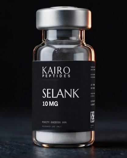
A gentle peptide, studied in research on mood regulation and focus.
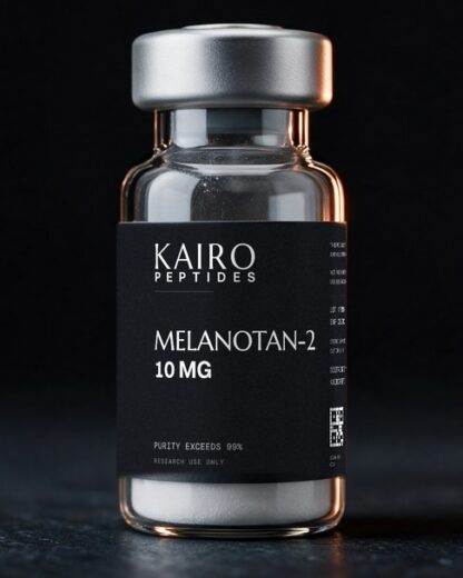
A melanocortin peptide, studied in research on skin pigmentation and erectile response.
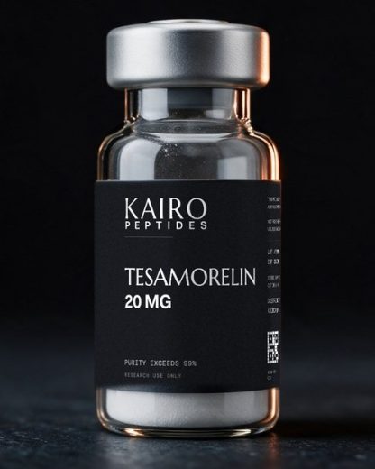
A well-documented peptide, studied in research on growth hormone release.